
https://www.youtube.com/embed/qhRwr0XRmBM?rel=0
I am Dr. Kevin Herman, a vascular and interventional radiologist at American Endovascular and Amputation Prevention. Today, we are going to talk about a Critical Limb Ischemia (CLI) case overview showing how we save limbs that others cannot.
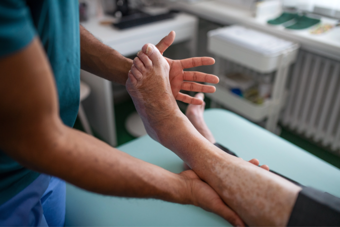
Patient history and consultation.
This is a 54-year-old male, presenting with a painful non-healing wound prior to an amputation site of the second and third digits of the right foot. The wounds have been present for more than three months prior to the initial consultation for a second opinion at our New Jersey Endovascular Center. His past medical history includes end-stage renal disease on hemodialysis, hypertension, and dyslipidemia. The patient was also a prior smoker.
His medications include:
- Metoprolol
- Novolin
- Lopressor
- Omeprazole
- Famotidine
- Atorvastatin
- Allopurinol
* Note that the patient was not on any antiplatelet therapy at that time.
Treatment Plan
The patient started with a diagnostic workup, which included:
- Lab evaluation, including evaluation for hemoglobin A1c, if not done recently.
- We would begin the patient on antiplatelet therapy.
- As well as perform arterial imaging and arterial Doppler studies.
- With that, we would then proceed to possible angiography and plan revascularization.
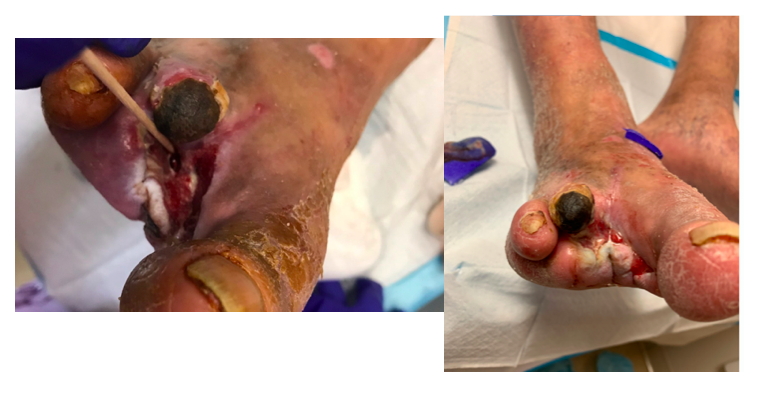
Here’s a clinical image of the initial presentation in March of 2020. As you can see, able to probe to the bone and the non-healing wound at the second and third amputation site.
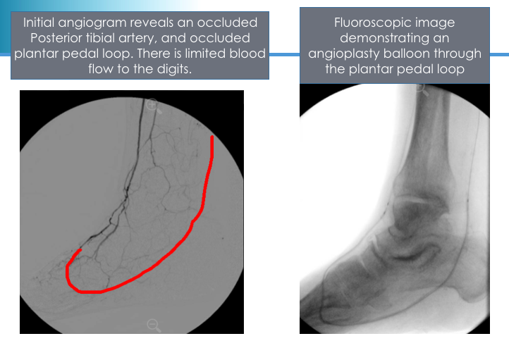
The initial angiogram revealed an occluded posterior tibial artery, as well as an occluded plantar pedal loop with limited blood flow to the digits. The graphic in red denotes where the normal posterior tibial artery and the plantar pedal loop would reside. A fluoroscopic image, after successful passing of the wire through the posterior tibial artery and plantar pedal loop, is shown here with an angioplasty balloon going through the plantar pedal loop and into the anterior tibial artery circulation.
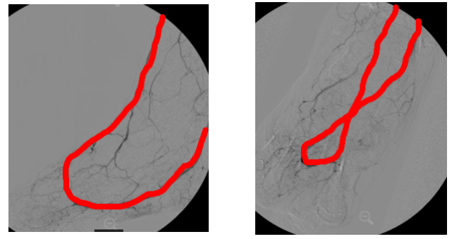
After the prolonged balloon angioplasty, a final angiogram was performed, and both seen in the lateral and AP projection showing the plantar pedal loop and posterior tibial artery, with a significantly improved flow to the pedal vessels. You can see, based on these graphic images, the anterior tibial artery to plantar pedal loop and posterior tibial circulation is now patent, as well as significantly improved flow to the pedal vessels and specifically at the site of amputation.
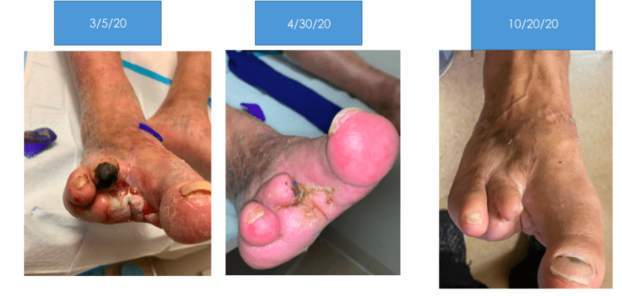
Photographic image of the patient follow-up with an initial presentation on the left, followed by a follow-up approximately one month, one-and-a-half months later, and then most recently with complete wound healing, and limb salvage and preservation in this patient with significant risk for limb loss.
Related Blogs & Videos
Learn more about vascular health, prevention, and care for Peripheral Artery Disease.


Request an Appointment at your nearest American Endovascular affiliated center.